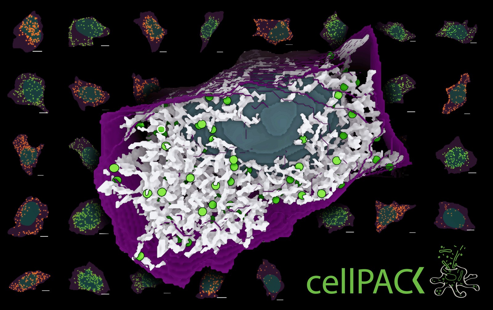
After a year of getting cellPACK back on its feet as a standalone Python package, we finally pointed it at a real biology question. Are peroxisomes (small, punctate organelles that detoxify molecules and break down fatty acids) randomly distributed in human iPS cells, or are they organized by some other rule?
The setup made use of our already published Allen Institute hiPSC dataset: ~305 segmented images of cells with endogenously tagged peroxisomes (PMP34), each one a snapshot of the spatial distribution we're trying to explain. We generated equivalent populations of simulated cells with cellPACK under three hypotheses: peroxisomes distributed randomly throughout the cytoplasm, biased toward the nucleus, or biased toward the plasma membrane. Each simulated cell is rendered as a multi-channel voxelized image — the same format as the experimental data — so the same downstream analysis pipeline can compare them on equal footing.
PILR, and a clear winner
We used PILR (Parameterized Intracellular Localization Reduction) to compute the average distribution profile for each population: real peroxisomes, random simulated, nuclear-biased simulated, membrane-biased simulated. We then computed correlations between each simulated profile and the experimental profile. The result was the nuclear-bias rule produced the highest correlation with the observed peroxisome distribution. Random and membrane bias correlated less well.
That's not a final answer — "nuclear bias" is a phenomenological description, not a mechanism and our rule still doesn't fully match the observed distribution. An abstract on this work was accepted for ASCB (we submitted to BPS too, awaiting decision). The team: Saurabh Mogre, Ruge Li, and me. We're working on extending to other punctate organelles like endosomes next, and getting the rule-mixing infrastructure ready for combined hypotheses.