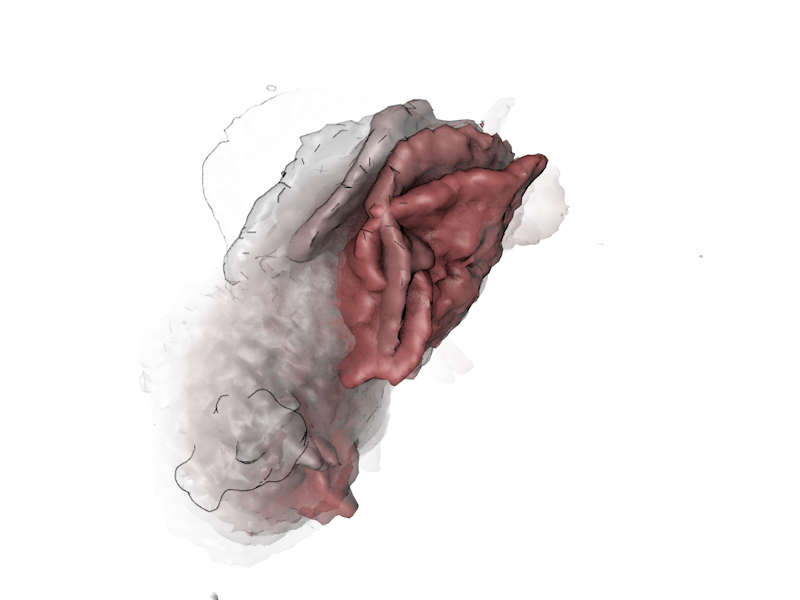
Lattice light sheet microscopy produces full 3D volumes over time. A typical migrating cell yields a 4D dataset — three spatial dimensions plus time — that a biologist needs to interpret. The native medium for that interpretation is still a 2D page. How do you compress 4D into 2D without losing what makes the cell legible as a moving object?
I took inspiration from artistic renderings of motion: 1. the cave at Chauvet, the 32,000-year-old bison drawings show extra legs, motion compressed into a single static figure, 2. Muybridge's 1878 horse-in-motion plates solved it differently: time made discrete, laid out in a strip, 3. Duchamp's Nude Descending a Staircase (1912) does both — superimposed phases of motion in a single composition. Each is an answer to the question of how a static image can encode change.
What I borrowed
For the HL60 migrating-cell work in the Mullins lab, I tried three encodings. First: classical frame strips at 18.8-second intervals — the Muybridge solution — for cell shape over time. Second: time-coded volumetric overlays where each color represents a 4-second window of protrusion activity, so an entire cycle of cell extension reads as a single Duchamp-like figure. Third: 3D-printed cells from the volumetric data, mounted on a hand crank for the Exploratorium so visitors could turn the time axis themselves. Each one trades different things: legibility for completeness, single-glance reading for full reconstruction.
The lesson is that the visualization problem for live cell imaging isn't a software problem — Chimera, Imaris, FIJI all render 4D fine. It's a representational problem, and there's a long lineage of solutions outside biology that I think we under-borrow from.