LDL & HDL Models
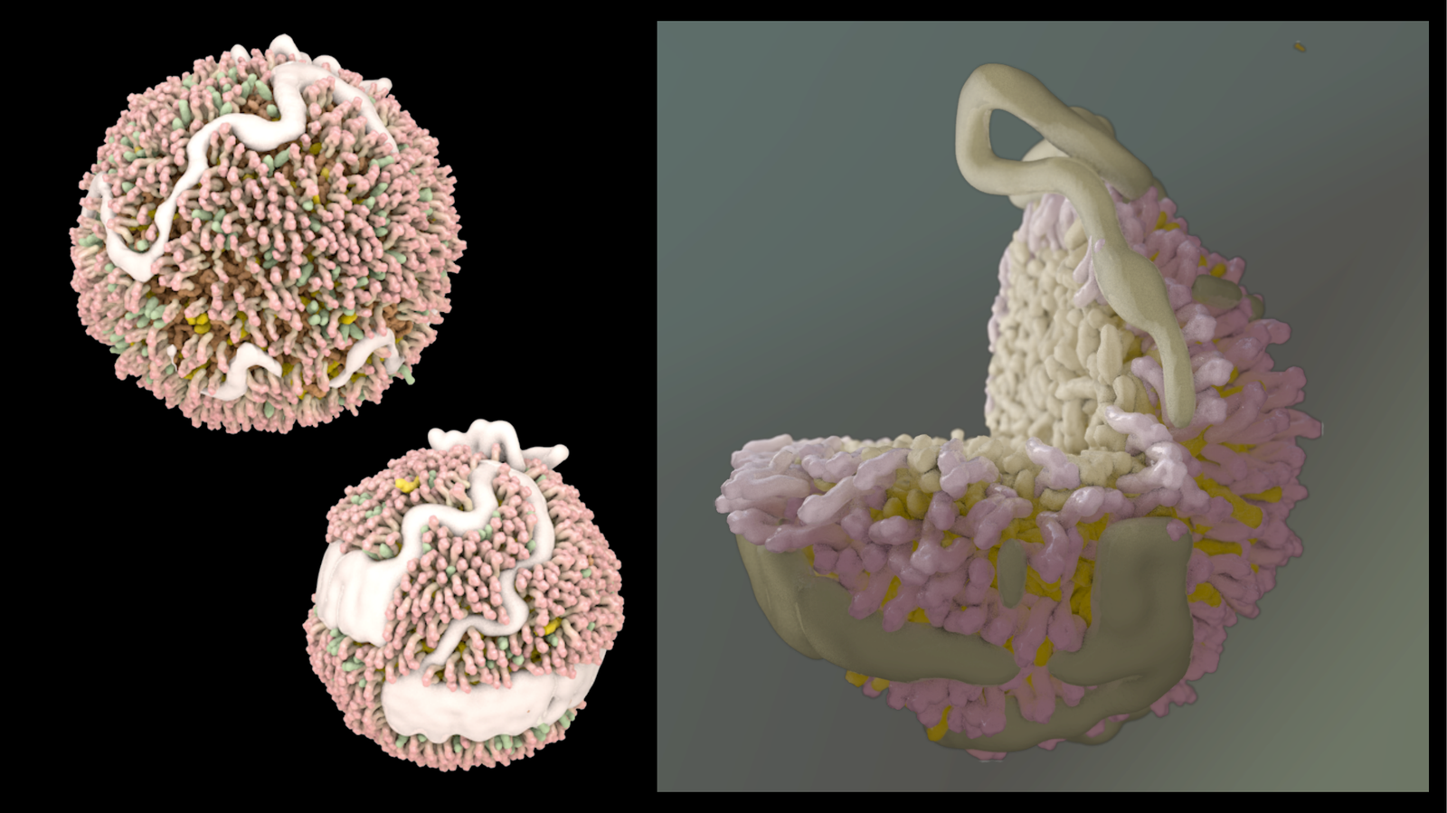
Detailed molecular models of low-density and high-density lipoprotein particles — built from structural data for use in education, research presentations, and public communication.
LDL and HDL are among the most clinically discussed molecules in medicine and among the least accurately depicted. Most popular illustrations are schematic at best: a colored ball with a few proteins dotted on the surface. These models were built to be structurally faithful — using published data on the lipid composition, apolipoprotein arrangement, and particle geometry — at a level of detail meant to hold up to scrutiny from a structural biologist.
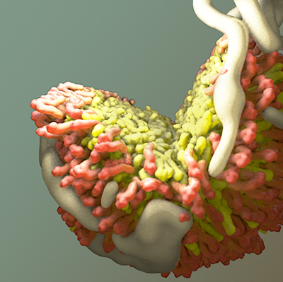
Most people have heard of these molecules. Almost no one has seen them accurately.
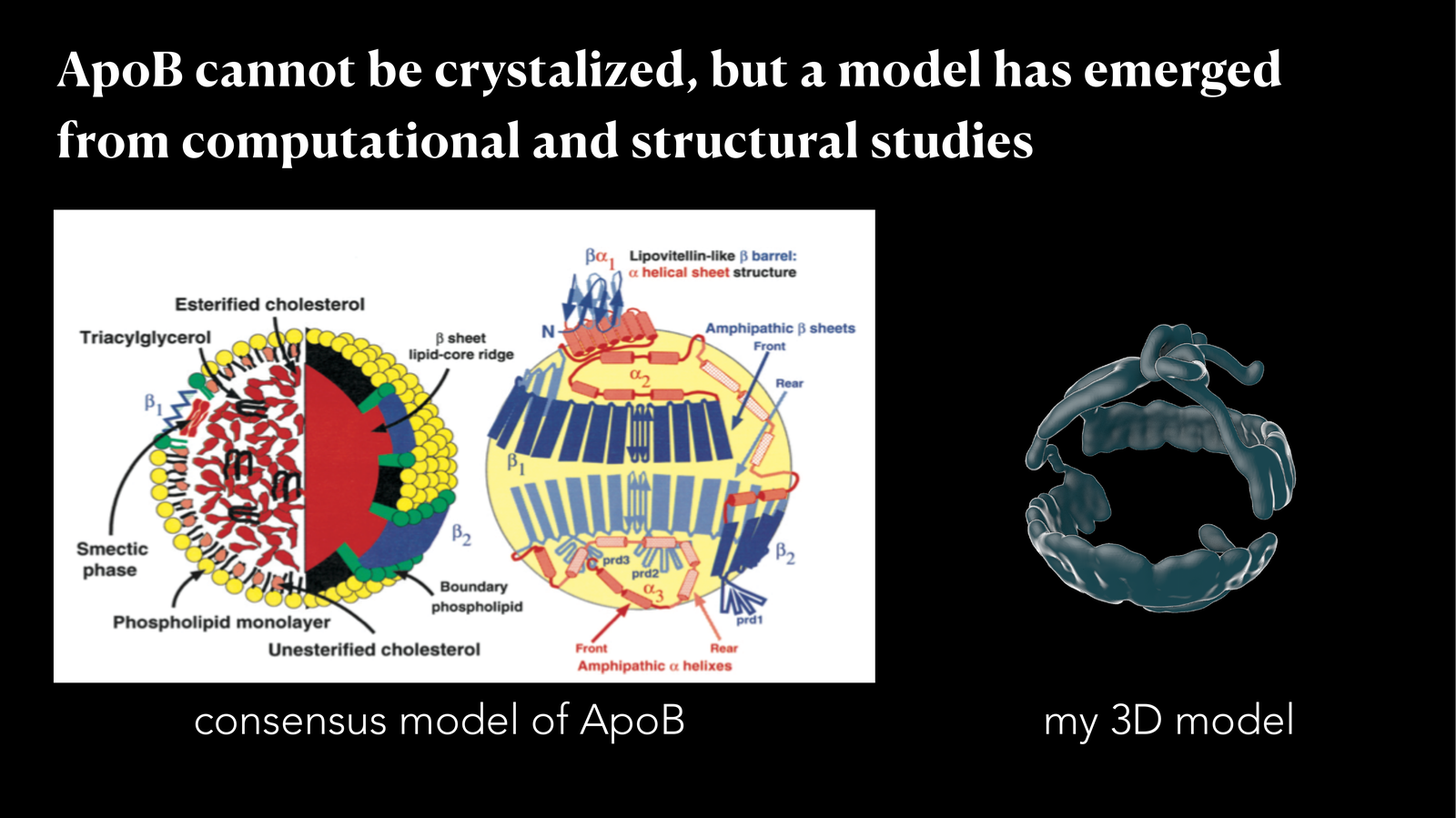
The hard part is Apolipoprotein B-100, the single enormous protein that wraps each LDL particle. ApoB cannot be crystallized — it is too large, too flexible, too embedded in lipid — so no atomic structure exists. What does exist is a consensus model assembled from decades of biochemistry, cryo-EM, and computational work: a lipovitellin-like β-barrel domain anchoring amphipathic α-helices and β-sheets across the particle surface. I worked from that consensus, building a 3D model in Cinema 4D that translates the schematic into space — proportions, surface texture, and the geometry of how the protein actually wraps the lipid core.
The models were used in research presentations and educational materials. Building them deepened my appreciation for how much scientific illustration still relies on convention rather than data — and how productive it can be to ask, for a molecule everyone draws schematically, what it would actually look like.